TII’s Vision: Leveraging Eye Images for Medical Innovations

At least 1 billion people worldwide have impaired vision that could have been prevented or has gone untreated, according to the World Health Organization.
Among the leading causes are cataracts, age-related macular degeneration, glaucoma, myopia, diabetic retinopathy, and other diseases.
Eric Buckland believes there’s a better way to study these eye disorders and to expedite the development of new diagnostics and therapies. His new medical software company, Translational Imaging Innovations (TII), is developing tools to help scientists and doctors get medical innovations into the eye clinic faster, with less frustration and at a lower cost.
“We are multimodal experts in the imaging of the eye,” said Buckland, an entrepreneur and Ph.D. scientist with 30 years of experience in optical technologies. “We have expertise in the imaging hardware and the image processing techniques, and we combine that with expertise on the clinical research workflows and the regulatory requirements.”
Photoreceptor images are key

To study eye disorders, scientists rely heavily on images of photoreceptors, the specialized rod and cone cells in the retina of the eye that respond to light, making vision possible.
How these cells are distributed in the retina can provide vital biomarkers for eye diseases. In fact, researchers typically use 10 different metrics to describe the distribution patterns of photoreceptors, Buckland said.
Many technologies are used to image the eye, but there is no systematic way for scientists and clinicians to process those non-standard images in large volumes.
“There’s a lack of interoperability so there’s a problem with how we use all of the data to extract medical insights,” Buckland said.
His company is the first to develop a software and analytics workflow system to solve that problem.
“We need a different way to think about how we collect, manage and process images so that we can translate the medical insight that we can derive from the imaging of the eye to clinical diagnostics and treatments, using biomarkers that we can derive from all these images,” Buckland said.
A three-part solution
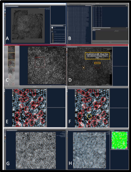
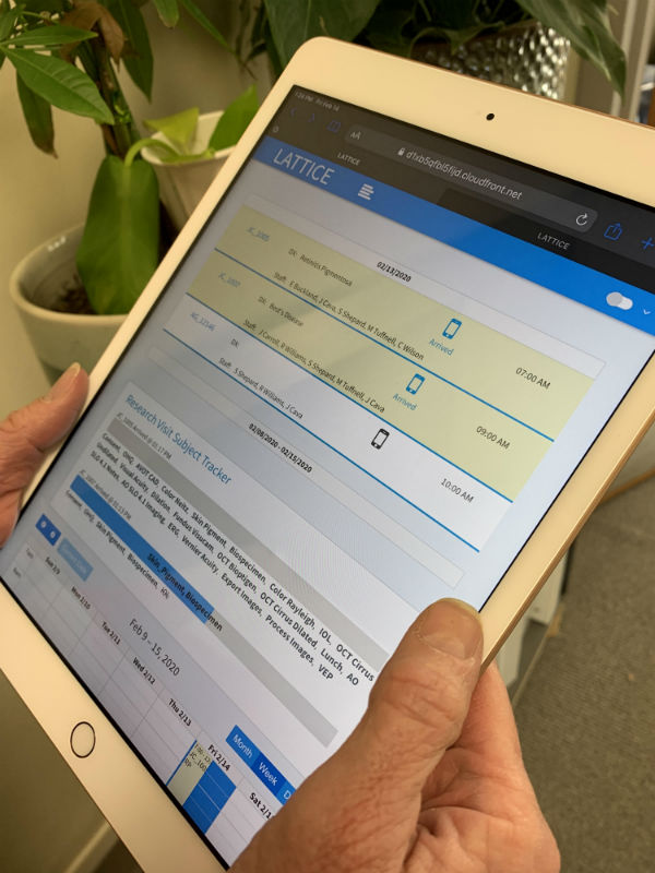
TII’s system consists of three parts: Lattice, a cloud-based software for clinical research workflow and data collection in both human and animal studies; Mosaic, an image-processing platform; and data from eye images.

“We’re stitching these three pieces -- Lattice to data, data to Mosaic, and Mosaic to Lattice -- into one single platform,” Buckland said. “Our target is to have a fully integrated system by the end of 2021. Until then we will be slowly developing the market with lead adopters.”
Lattice was licensed from its developer, the Medical College of Wisconsin’s Advanced Ocular Imaging Program. In the last two years Lattice has housed data from about 3,000 research patients and 80 research protocols. It will soon be deployed at Tier 1 research institutions in the Northeast, the West Coast and the British Isles, Buckland said.
Mosaic was developed by Robert Cooper, Ph.D., now assistant professor of biomedical engineering at Marquette University, when he was a doctoral student. It is in use at about six institutions including the Foundation Fighting Blindness, a Raleigh-based nonprofit organization that is the world’s largest private funder of retinal disease research. The Foundation is using the analytics tool to study Usher syndrome, an inherited eye disease.
When the system is further developed and integrated, TII’s first paying customers will be academic researchers who are either developing new analysis techniques to diagnose or predict disease progression, or using imaging systems to understand disease or treatment in small animals or in the clinic, Buckland said.
The next tier of customers will be pharmaceutical companies that are developing treatments for eye diseases or neurological disorders involving the retina.
“The demand from pharma to have new outcome measures and new end points for their (drug) development is very clear, Buckland said.
“The other trajectory that’s happening is that with artificial intelligence (AI) there will be more tools to help clinicians make decisions using guidance from AI type of algorithms. We see us growing from research to therapy development to clinical management in the long term.”
Major research funding
Earlier this year TII received a $1.5 million Direct-to-Phase II Small Business Innovation Research (SBIR) award from the National Eye Institute of the National Institutes of Health. The grant will allow the company to use its Mosaics platform to analyze photoreceptor patterning in images of 1,800 patients afflicted with degenerative eye disease.
“It is critical to leverage the untapped medical information hidden within these images to advance diagnosis and accelerate the development of new therapies to treat blinding eye disease,” Buckland said. “This program is the beginning of our mission to unleash the power of the eye and transform medicine.”
The images were acquired by clinical researchers at Medical College of Wisconsin over a decade of research into inherited retinal disease. Renowned eye researcher Joseph Carroll, Ph.D., professor of ophthalmology and visual sciences and director of the Advanced Ocular Imaging Program, is a co-principal investigator on the project with Buckland.
“Adaptive optics imaging has emerged as a sensitive marker of the presence and viability of photoreceptors,” Carroll said. “However, there are no validated algorithms or objective quantitative measures based on adaptive optics images of the photoreceptor mosaic. A representative image database supported by quantitative metrics is necessary to screen patients for treatment and manage follow-up.”
A return to the Triangle

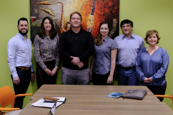
The TII team (from left): Andrew Witchger, senior software engineer; Maureen Tuffnell, product manager;
Rob Williams, chief technical officer; Taylor Szeto, product engineer; Eric Buckland, CEO;
and Tammy Carrea, vice president of regulatory and clinical affairs.
For now, TII is a virtual company with CEO Buckland and five employees working from their homes. Buckland said he plans to relocate the company from his home in Hickory to the Research Triangle area this year.
Prior to starting TII, Buckland was founder and CEO of Bioptigen, a 2004 spinout of Duke University that provided eye-imaging systems for translational research. The company was sold in 2015 to Leica Microsystems, a German-based global leader in microscopy and imaging systems. Buckland stayed on as Leica’s general manager and senior director in Morrisville until 2017.
He founded TII when he realized that many age-related eye disorders were becoming more common and more acute as populations aged. The time seemed right to develop a better way of processing and analyzing photoreceptor images, drawing on new tools of big data and artificial intelligence.
“The most interesting innovations occur where there’s a convergence of multiple trends,” he said.
